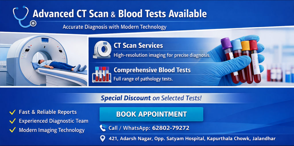
Services
50-Slice CT Scan & Angiography
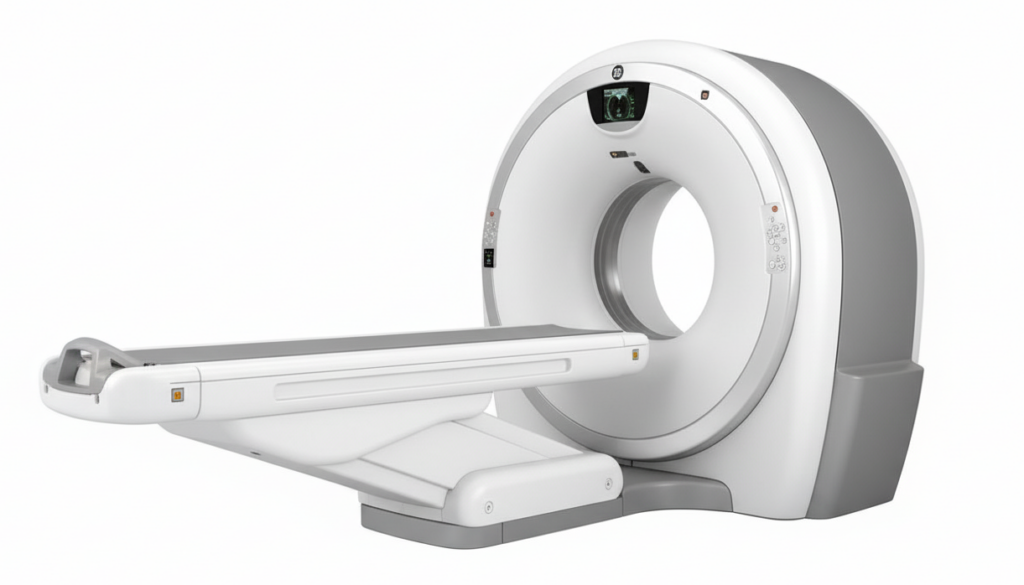
Alfa Imaging & Diagnostic Labs pioneered advanced 50-slice CT technology in the region, offering high-precision 3D reconstruction and isometric imaging for comprehensive diagnosis. Our cutting-edge scanners deliver exceptional clarity at routine CT scan prices.
NABH Accredited
PC-PNDT Compliant
50-Slice CT Scanner
Ultra-thin 0.625mm slices for unparalleled detail
Advanced Angiography
Pulmonary, brain, and vascular studies
Virtual Procedures
Colonoscopy & bronchoscopy without scopes
Pressure Injector
Precision contrast delivery for enhanced imaging
Advanced CT Technology
Our 50-slice CT scanner provides ultra-thin 0.625mm slices with volume acquisition and isometric reconstruction in any plane. This revolutionary technology enables:
- True-match imaging for precise diagnostics
- Whole-body scanning capabilities
- Reduced radiation exposure
Despite these advanced capabilities, patients pay standard CT scan rates, making high-quality diagnostics accessible to all.
About
Pressure (Contrast) Injectors
A pressure injector (or power injector) is a medical device that automatically delivers contrast agents at precise, high flow rates and pressures for diagnostic imaging (like CT, MRI) and interventional procedures, ensuring clear visualization of blood vessels and organs, with dual-head models also flushing saline.
- •Function: Delivers contrast dye into a patient's bloodstream for CT, MRI, and angiography to highlight blood vessels and abnormalities.
- Mechanism: Uses motor-driven plungers to push contrast from syringes at controlled speeds and pressures, often exceeding manual capabilities.
- Components: Includes an injector head, syringes, and tubing, often with a saline flush feature (dual-head).
- Applications: CT angiography, cardiac CT/MRI, perfusion imaging, and cardiac catheterization.
What it Shows
- Blood Flow: Detailed images of arteries (carotid, vertebral) and veins.
- Vascular Abnormalities: Aneurysms (bulges), narrowing, blockages (stenosis), or tears (dissections).
- Brain Supply: Vessels feeding the brain, including the aortic arch, carotids, and vertebrals.
Angiography Services
CT Renal Angiogram
A CT Renal Angiogram (CTA) is a specialized X-ray test using contrast dye to create detailed 3D images of the arteries and veins supplying the kidneys, helping diagnose blockages (stenosis), bulges (aneurysms), or other vascular issues causing high blood pressure, kidney pain, or failure. It's a fast, non-invasive tool that helps doctors see blood flow, plan surgeries, and manage kidney-related vascular problems.
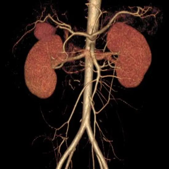
What it's used for (Indications)
- Renal Artery Stenosis: Narrowing of kidney arteries, a common cause of severe hypertension.
- Aneurysms: Bulging or weakened areas in the artery wall.
- Blockages/Thrombosis: Clots or obstructions in renal arteries or veins.
- Vascular Malformations: Abnormal connections between blood vessels.
- Kidney Tumors: To assess involvement of blood vessels.
- Pre-Surgical Planning: For kidney transplants or surgeries.
- Unexplained Kidney Injury/Failure: To find the underlying vascular cause.
What to expect during the procedure
Preparation: May involve fasting and blood tests.
During Scan: You lie on a table; contrast dye is injected, causing a brief warm sensation.
Post-Scan: Drink plenty of fluids to flush the contrast;certain diabetic medications (like Metformin) may need to be paused.
Benefits
- • Provides detailed, high-resolution images.
- • Helps diagnose conditions early, often before severe symptoms appear.
- • Increasingly replacing older, more invasive methods like conventional angiography.
Head and Neck (Brain) Angiogram
A Head and Neck CT Angiography (CTA) is a specialized, non-invasive imaging test using X-rays and a contrast dye to create detailed 3D pictures of blood vessels in the head and neck, diagnosing issues like aneurysms, blockages (stenosis), dissections, or vascular malformations in arteries like the carotid and vertebral arteries that supply the brain, helping to find stroke causes, tumors, or trauma-related damage. The procedure involves lying in a scanner, receiving an iodine-based dye via IV, and holding still as images are taken to visualize blood flow, with quick scans capturing the dye's path.
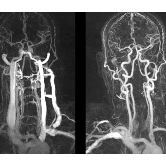
Why it's Done (Symptoms & Conditions)
- Stroke Symptoms: Sudden weakness, numbness, speech/vision problems, dizziness, loss of balance, severe headache
- Stroke Evaluation: Identifying the cause, such as blockage or dissection.
- Trauma: Assessing vascular injury after head/neck trauma.
- Tumors: Checking blood flow patterns to guide treatment.
The Procedure
- Preparation: An IV line is placed in your arm.
- Contrast Injection: A special contrast dye (iodine-based) is injected, often causing a warm feeling.
- Scanning: You lie on a table, head-first into the CT scanner.
- Imaging: The scanner takes rapid images as the dye moves through vessels.
- Image Reconstruction: Computer software creates detailed 3D images
Perfusion CT (Computed Tomography) scan
A Perfusion CT (Computed Tomography) scan is a functional imaging technique that uses rapid X-ray scans after contrast injection to create maps showing blood flow (CBF), blood volume (CBV), and time (MTT) in tissues, especially the brain, helping diagnose strokes by identifying salvageable tissue (penumbra) from dead tissue, guiding treatment like thrombolysis, and assessing tumors or vasospasm. It provides real-time physiological data, unlike standard CT, to assess tissue viability and treatment effectiveness quickly
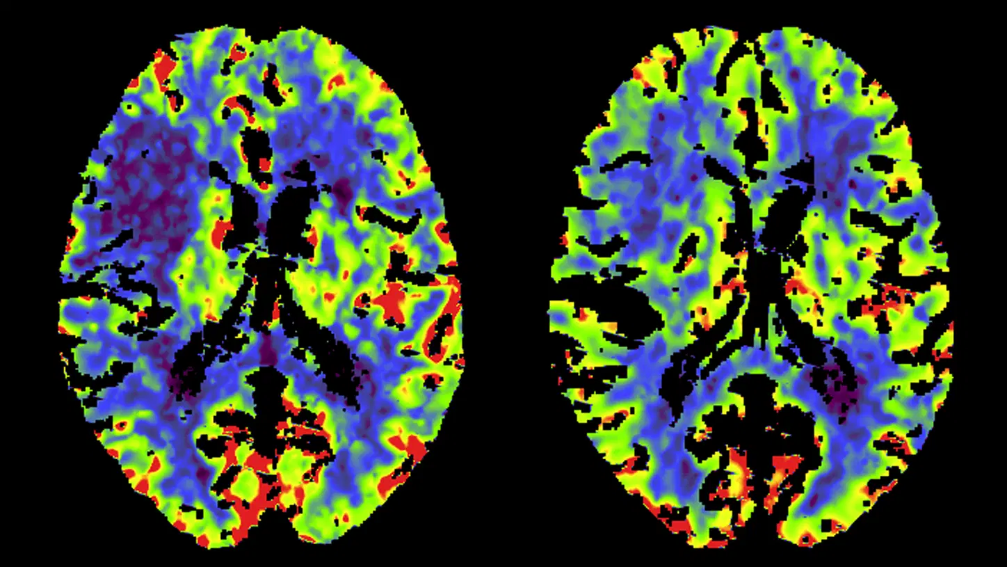
How it Works
- Contrast Injection: A contrast dye is rapidly injected into a vein.
- Rapid Scanning: A CT scanner takes many images in quick succession as the contrast travels through the blood vessels.
- Mapping: Specialized software analyzes these images to generate color-coded maps of blood flow, volume, and transit time.
Key Clinical Uses (Primarily Brain)
- Acute Stroke: Differentiates the core infarct (dead tissue) from the ischemic penumbra (tissue at risk but potentially salvageable).
- Treatment Guidance: Helps decide if a patient needs clot-busting drugs (thrombolysis) or clot retrieval for stroke.
- Vasospasm: Monitors blood flow in conditions like subarachnoid hemorrhage.
- Brain Tumors: Assesses tumor vascularity and treatment response.
- Cerebrovascular Reserve: Identifies patients who might benefit from bypass surgery.
Benefits
- Speed: Provides crucial information rapidly, vital in stroke emergencies.
- Non-Invasive: A painless test performed with standard CT equipment.
- Functional Data: Shows how the brain is working, not just its structure.
CT Pulmonary Angiography
A CT Pulmonary Angiography (CTPA) is a specialized CT scan using contrast dye to visualize the blood vessels in the lungs, primarily to diagnose pulmonary embolism(blood clots), but also to assess pulmonary hypertension, AV malformations, or congenital vessel narrowing. The non-invasive procedure involves an IV injection of contrast, with patients holding their breath for short scans, revealing blockages or other abnormalities as filling defects in the arteries.
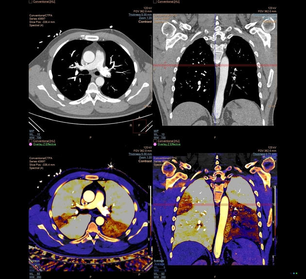
What it's used for:
- Pulmonary Embolism (PE): The main reason for the test, detecting acute or chronic blood clots.
- Pulmonary Hypertension: High blood pressure in the lung arteries.
- Arteriovenous Malformations (AVMs): Abnormal connections between arteries and veins in the lungs.
- Congenital Abnormalities: Narrowing or other birth defects in pulmonary vessels.
- Pulmonary Artery Aneurysms: Bulging of the artery walls.
How it works:
- Preparation: You’ll lie on your back on a CT table, often changing into a gown, and have an IV placed in your arm or hand.
- Contrast Injection: A contrast dye is injected through the IV, which travels to the lungs’ blood vessels.
- Scanning: The CT scanner takes rapid images as the contrast highlights the vessels. You’ll be asked to hold your breath briefly during scans.
- Analysis: Radiologists look for “filling defects” (areas where the contrast doesn’t fill the vessel) to identify clots or other issues.
What to expect:
- Sensation: A brief flushing, salty taste, or slight headache might occur when the contrast is injected.
- Duration: The scanning part is quick, often around 10 minutes.
- Non-Invasive: It's a non-invasive test, meaning no surgery is involved, though an IV is used.
Upper Limb CT Angiography (CTA)
Upper Limb CT Angiography (CTA) is a fast, minimally invasive imaging test using CT scans and contrast dye to create detailed pictures of arteries in the arm, shoulder, forearm, and hand, crucial for diagnosing blockages (like from plaques or clots), trauma, infections, and planning surgeries by showing vessel health and surrounding tissues. It's a valuable tool for acute situations like limb ischemia or injury, offering quick, non-invasive assessment compared to older methods like DSA, though digital subtraction angiography (DSA) might still be better for fine details in the hand
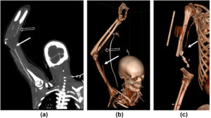
What It Is & How It Works
- Technology: Combines a CT scanner with intravenous (IV) contrast dye to highlight blood vessels.
- Procedure: You lie on a CT table, have an IV placed (often in the arm), and the scan captures images as dye flows through your arm arteries.
- Speed & Detail: It's quick, often taking minutes, and provides detailed 3D views of arteries, showing narrowing, blockages, or injuries.
Why It's Done (Indications)
- Trauma: Assessing vascular damage from injuries.
- Ischemia: Diagnosing lack of blood flow (e.g., sudden coldness, pain).
- Infections: Identifying complications like infectious pseudoaneurysms.
- Vascular Abnormalities: Detecting aneurysms, narrowing (stenosis), or congenital issues.
- Pre-surgical Planning: Mapping vessels before complex reconstruction.
Advantages
- Fast & Available: Rapid acquisition, often 24/7 in emergencies.
- Minimally Invasive: Less invasive than traditional angiography.
- Shows Surrounding Tissues: Reveals both vascular and musculoskeletal structures, helpful for trauma.
Key Considerations
- Contrast Dye: Involves radiation and contrast, so pregnancy and kidney issues need to be disclosed.
Lower Limb CT Angiography (CTA)
Lower Limb CT Angiography (CTA) is a fast, minimally invasive imaging test using CT scans and contrast dye to create detailed pictures of arteries in the arm, shoulder, forearm, and hand, crucial for diagnosing blockages (like from plaques or clots), trauma, infections, and planning surgeries by showing vessel health and surrounding tissues. It's a valuable tool for acute situations like limb ischemia or injury, offering quick, non-invasive assessment compared to older methods like DSA, though digital subtraction angiography (DSA) might still be better for fine details in the leg.
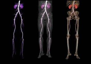
What It Is & How It Works
- Technology: Combines a CT scanner with intravenous (IV) contrast dye to highlight blood vessels.
- Procedure: You lie on a CT table, have an IV placed (often in the arm), and the scan captures images as dye flows through your arm arteries.
- Speed & Detail: It's quick, often taking minutes, and provides detailed 3D views of arteries, showing narrowing, blockages, or injuries.
Why It's Done (Indications)
- Trauma: Assessing vascular damage from injuries.
- Ischemia: Diagnosing lack of blood flow (e.g., sudden coldness, pain).
- Infections: Identifying complications like infectious pseudoaneurysms.
- Vascular Abnormalities: Detecting aneurysms, narrowing (stenosis), or congenital issues.
- Pre-surgical Planning: Mapping vessels before complex reconstruction.
Advantages
- Fast & Available: Rapid acquisition, often 24/7 in emergencies.
- Minimally Invasive: Less invasive than traditional angiography.
- Shows Surrounding Tissues: Reveals both vascular and musculoskeletal structures, helpful for trauma.
Key Considerations
- Contrast Dye: Involves radiation and contrast, so pregnancy and kidney issues need to be disclosed.