Services
Fetus Scan
Pregnancy ultrasound - Schedule and importance of dating, growth, anomaly scan
Which | When | Why
By
Dr. Mayank Gupta
M.B.B.S., M.D Radiodiagnosis (FMF , UK Certified) Ex Sr. Residency BVP-Pune , Rajiv Gandhi Cancer Hospital, New Delhi Ex Associate Consultant, Sir Ganga Ram Hospital, Delhi Society of Foetal Medicine IRIA, IMA, International Society of Ultrasound in Obstetrics and Gynecology (ISUOG)
Words of motivation
As a radiologist, every pregnancy scan tells a beautiful story of new life unfolding. When an expectant mother gently rests her hands on her belly, we see more than just images on a screen—we witness a powerful bond forming between mother and child. Each heartbeat, each movement captured during the ultrasound reflects the miracle of growth happening inside her womb. Pregnancy is a remarkable nine-month journey, where every stage brings new developments. From the first tiny heartbeat to the well-formed features of the baby, our role is to carefully monitor this progress and ensure everything is developing healthily. With advanced imaging and precise diagnostics, we help mothers feel confident and reassured at every step. Our commitment is to provide accurate, compassionate care so that every mother can enjoy this phase without worry. Because when it comes to nurturing new life, nothing matters more than trust, expertise, and the right medical support.
NABH Accredited
PC-PNDT Compliant
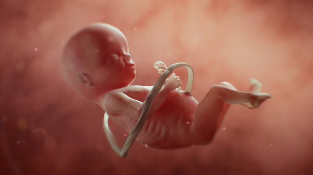
Pregnancy is a nine-month journey during which a baby gradually develops from a few cells into a fully formed human being. Regular ultrasound scans play a vital role in monitoring this growth and ensuring the health of both mother and baby. Key scans such as the dating scan, anomaly scan, cardiac scan, and growth scan are performed at specific stages of pregnancy to assess development, detect abnormalities, and track overall well-being. When done on time, these specialized ultrasounds provide essential medical information to support safe and healthy motherhood.
Pregnancy is divided into three trimesters, each lasting three months. During this time, the baby develops gradually, making regular monitoring essential. As the mother’s body undergoes many changes, timely doctor visits, proper nutrition, medications, and scheduled ultrasound scans help ensure a safe, smooth, and healthy pregnancy journey.
There are some ultrasounds scan that needs to be performed during one's period of pregnancy. We shall have a closer look at the most significant and necessary ultrasounds offered in one's pregnancy from the first to third trimesters. Here is a quick summary of the pregnancy ultrasound scan.
| Trimester | Week | Scan name | Purpose |
|---|---|---|---|
| 1st | 6 to 8 | Early pregnancy or dating scan | Confirm the pregnancy, its position, and estimate the baby’s age. |
| 11 to 13 | NT / ND scan | Measures Nuchal Translucency, check for nasal bone, examine the spinal column. Uterine artery doppler is also checked. | |
| 2nd | 18 to 20 | Anomaly or TIFFA scan | Check anatomic structures and find out a defect in the baby and placenta. Most important pregnancy scan. |
| 24 to 26 | Cardiac scan / Fetal echocardiography | Evaluate structural and functional integrity of fetal heart. | |
| 3rd | 28 to 32 | Pregnancy welfare and growth scan | Check the progress of the baby’s growth and status of liquor (fluid around the baby). |
| 28 to 32 | Obstetric doppler scan (Optional) | Measures the blood flow through the umbilical cord to the different internal organs of the developing baby’s body | |
| 36 to 40 | Growth scan. Obstetric doppler scan (If needed) | Check baby’s growth, position, amount of liquor, and blood flow to the baby. |
First Trimester Ultrasound

Pregnancy is a beautiful journey in which a baby gradually develops day by day inside the womb. During the early weeks, the mother plays a vital role in nurturing both herself and the growing life within her. By 6 to 8 weeks of pregnancy, the developing cells form a small body mass, and important physical changes become visible.
Two essential ultrasound scans are recommended during the first trimester. The first is the dating scan (6–8 weeks), which confirms pregnancy, detects the baby’s heartbeat, determines gestational age, and checks for abnormalities such as internal bleeding or ectopic pregnancy. It also helps identify twin or multiple pregnancies. This scan is usually performed using a transvaginal approach for clearer imaging, though an abdominal method may also be used.
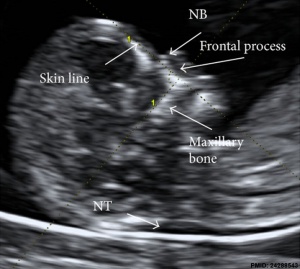
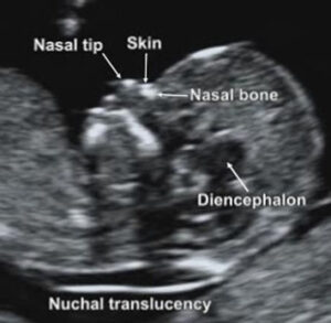
The second important scan is the Nuchal Translucency (NT) scan, done between 11 weeks and 13 weeks 6 days. This scan measures the fluid at the back of the baby’s neck and checks spinal development and the presence of the nasal bone. It is commonly combined with a double marker blood test to assess the risk of chromosomal abnormalities.
For twin pregnancies, the NT scan helps determine the type of twins and guides further care. Additional measurements such as uterine artery Doppler assess the risk of high blood pressure and growth issues, while cervical length evaluation helps identify the risk of preterm birth.
What is anomaly scan or TIFFA scan


What is best time to schedule and perform anomaly scan or TIFFA scan?
Importance of anomaly scan or TIFFA scan and why to go for it?
What baby details can be found from an Anomaly scan or TIFFA scan?
- Anencephaly (98%)
- Gastroschisis (98%)
- Edwards’ Syndrome (Trisomy 18) (95%)
- Patau’s Syndrome (Trisomy 13) (95%)
- Bilateral Renal Agenesis (84%)
- Exomphalos (80%)
- Cleft Lip (75%)
- Open Spina Bifida (90%)
- Diaphragmatic Hernia (60%)
- Lethal Skeletal Dysplasia (60%)
- Down Syndrome (50%)
- Congenital Heart Disease (50%)
For abnormalities , click here…
How the anomaly scan or TIFFA scan is done?
Is it necessary to take the anomaly scan or TIFFA scan?
Is there any harmful effect of the anomaly scan or TIFFA scan on the baby?
When do I receive my anomaly scan report or TIFFA scan report?
What happens if any abnormality of the baby is detected in the report?
Second Trimester Ultrasound

Cardiac scan / Fetal 2D echocardiography
Cardiac pregnancy scan is meant to check the structure and function of a baby’s heart. This scan examines how blood flows through your baby’s arteries and veins. This sonography is one of the optional sonographies during pregnancy. This pregnancy ultrasound scan is recommended typically in high-risk pregnancies.
This scan also looks out for abnormalities such as holes in the heart, narrowing of arteries, valves that don’t open and close properly. The cardiac scan is necessary for the following conditions:
- You were born with a heart defect
- You've already had a baby with a heart defect
- You have diabetes. This means you are at a slightly higher risk of having a baby with a heart defect
- You take certain drugs that can increase the risk of heart problems, such as some medications for epilepsy
- Your nuchal translucency (NT) scan shows that your baby has fluid at the back of her neck of 3.5mm or more but appears to have normal chromosomes


Third Trimester Ultrasound

Cardiac pregnancy scan is meant to check the structure and function of a baby’s heart. This scan examines how blood flows through your baby’s arteries and veins. This sonography is one of the optional sonographies during pregnancy. This pregnancy ultrasound scan is recommended typically in high-risk pregnancies.
Late pregnancy welfare scan or third trimester growth scan
The third-trimester growth scan, also known as the late pregnancy welfare scan, is usually performed between 28–32 weeks and again between 36–39 weeks of pregnancy. The number and frequency of these scans depend on the baby’s growth, health, and any medical concerns.
By this stage, the baby’s facial features and body structures are clearly visible, allowing better imaging. This scan evaluates whether the baby is growing properly by measuring weight, size, and other body parameters. It also confirms the baby’s position inside the womb.
Additionally, the scan assesses the placenta’s location to ensure it is not too close to the cervix and checks the level of amniotic fluid required for healthy development. Any signs of internal bleeding, discomfort, or conditions such as uterine fibroids are also examined.
In multiple pregnancies, such as twins or triplets, growth scans are especially important to monitor each baby’s development and detect any potential complications early.
Obstetric Doppler ultrasound (28-32 week ultrasound scan)
Proper blood flow to the uterus, placenta, and umbilical cord is essential for the healthy development of the baby during pregnancy. Any condition that restricts this blood supply can affect the baby’s growth and well-being, making regular monitoring important.
A Doppler ultrasound scan, usually performed between 28 to 32 weeks of pregnancy, is used to assess blood flow. Unlike a regular ultrasound, this scan shows how blood moves through the vessels. It helps detect any blockage or reduced circulation between the placenta and the baby.
This scan measures blood flow through the umbilical cord and to the baby’s vital organs, ensuring the baby receives sufficient oxygen and nutrients for proper growth.


Are all the pregnancy ultrasounds safe?
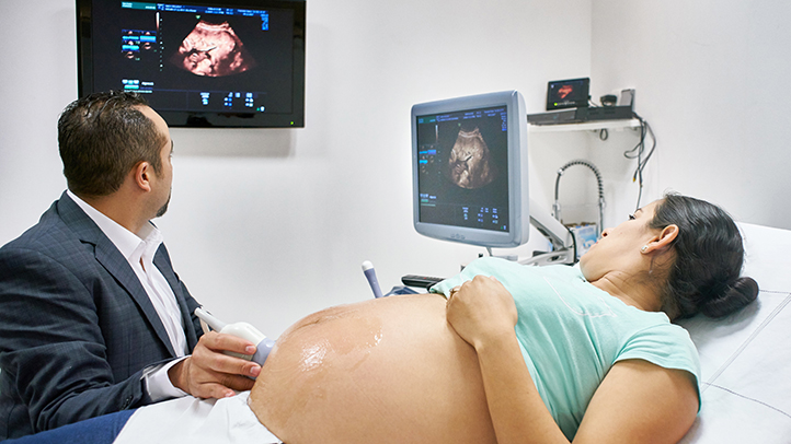
Pregnancy ultrasound scans are completely safe, as they use sound waves rather than radiation. These scans are performed by trained professionals such as sonographers, radiologists,. Conducted at different stages of pregnancy, they provide valuable information about the health and well-being of both the mother and the unborn baby.
Who performs the ultrasounds?
Skilled and trained medical professionals (Radiologist) perform these pregnancy ultrasound scan with utmost care and proficiency. With these dating scan and growth scan, they can understand the overall age and health of the baby.
Where can one get these pregnancy ultrasound scan done?
The pregnancy ultrasound scan are performed in various hospitals. There are also specialized labs that can perform these pregnancy scan. But it is imperative to talk and discuss it with your Radiologist to guide you to selecting the correct scan. Your gynecologist’s prescription is mandatory to do any pregnancy ultrasound scan.
Pregnancy ultrasound scan schedule summary
| Trimester | Week | Scan name | Purpose |
|---|---|---|---|
| 1st | 6 to 8 | Early pregnancy or dating scan | Confirm the pregnancy, its position, and estimate the baby’s age. |
| 11 to 13 | NT / ND scan | Measures Nuchal Translucency, check for nasal bone, examine the spinal column. Uterine artery doppler is also checked. | |
| 2nd | 18 to 20 | Anomaly or TIFFA scan | Check anatomic structures and find out a defect in the baby and placenta. Most important pregnancy scan. |
| 24 to 26 | Cardiac scan / Fetal echocardiography | Evaluate structural and functional integrity of fetal heart. | |
| 3rd | 28 to 32 | Pregnancy welfare and growth scan | Check the progress of the baby’s growth and status of liquor (fluid around the baby). |
| 28 to 32 | Obstetric doppler scan (Optional) | Measures the blood flow through the umbilical cord to the different internal organs of the developing baby’s body | |
| 36 to 40 | Growth scan. Obstetric doppler scan (If needed) | Check baby’s growth, position, amount of liquor, and blood flow to the baby. |
This article explains the most important pregnancy ultrasound scans, including the dating scan, anomaly scan, and growth scan, which are routinely recommended during pregnancy. Throughout the nine-month journey, a mother plays a vital role in nurturing both herself and her unborn baby. Regular doctor visits, prescribed medications, ultrasounds, a healthy diet, and a balanced lifestyle are essential parts of prenatal care.
In a normal pregnancy, these standard scans are usually sufficient. However, in special cases such as twin or triplet pregnancies, fetal growth concerns, or high-risk conditions, more frequent ultrasounds may be advised by the gynecologist. These scans help closely monitor the baby’s development and ensure timely medical support.
Watching your baby grow through each stage of pregnancy is a truly emotional and memorable experience, from a tiny group of cells to a fully formed life inside the womb.

M.B.B.S., M.D Radiodiagnosis (FMF , UK Certified) Ex Sr. Residency BVP-Pune , Rajiv Gandhi Cancer Hospital, New Delhi Ex Associate Consultant, Sir Ganga Ram Hospital, Delhi Society of Foetal Medicine IRIA, IMA, International Society of Ultrasound in Obstetrics and Gynecology (ISUOG)
Disclaimer

Ovulation Period
Know the best ovulation days to get pregnant
Want to get pregnant? Want to know your best fertility days? Use this ovulation calculator to know your ovulation period, symptoms, and best ovulation days to get pregnant