Services
High Frequency Xray and Procedures
High-frequency X-ray imaging is a cornerstone of modern medical diagnostics.
High-frequency X-ray imaging is a cornerstone of modern medical diagnostics. It delivers rapid, accurate, and non-invasive insights into internal anatomy and pathology, supporting early disease detection and effective treatment planning. With techniques ranging from conventional radiography to advanced CT scans, high-frequency X-rays remain essential in patient care.
NABH Accredited
PC-PNDT Compliant
About
What Are High-Frequency X-Rays?
High-frequency X-rays are a type of electromagnetic radiation with very short wavelengths and high energy, capable of penetrating body tissues to produce detailed internal images. In medical diagnostics, X-rays are used to visualize bones, organs, and soft tissues quickly and non-invasively, helping clinicians detect fractures, abnormalities, infections, and other conditions with clarity.
How Do High-Frequency X-Rays Work?
When high-frequency X-rays pass through the body, different tissues absorb radiation at different rates. Dense structures like bones absorb more X-rays, appearing lighter on the resulting image, while softer tissues absorb less and appear darker. Specialized detectors capture the remaining X-rays and convert them into digital images that doctors can interpret for diagnosis and treatment planning.
Why Choose High-Frequency X-Ray Imaging?
Fast and Efficient: X-ray imaging is rapid and often completed in minutes, allowing for quick clinical decisions.
Non-Invasive: It does not require incisions or invasive tools, making it comfortable and safe for patients.
Wide Diagnostic Use: X-rays assist in evaluating bone health, chest conditions, abdominal issues, dental concerns, and more.
Real-Time Visualization: Techniques like fluoroscopy enable continuous internal imaging during procedures.
Detailed Internal Views: CT scans expand diagnostic ability with detailed cross-sectional imaging.
Safety and Radiation Considerations
X-rays use ionizing radiation, which can slightly increase long-term health risks at high doses. However, modern protocols and equipment minimize exposure by using the lowest radiation necessary to obtain quality images. Radiology teams also use shielding and protection measures when appropriate.
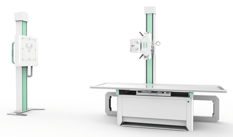
Types of X-Ray Imaging Procedures
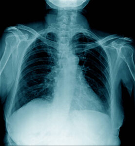
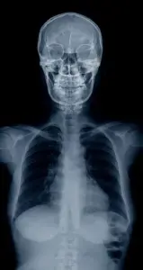
Conventional X-Ray (Radiography)
This is the most widely used X-ray technique. It captures a single still image of a specific body part, such as the chest, limbs, or spine, and is essential for diagnosing fractures, lung infections, arthritis, and other conditions.
Fluoroscopy
Fluoroscopy uses continuous X-ray beams to create real-time moving images. It is particularly useful during guided procedures — for example, to monitor the position of instruments, contrast flow through vessels, or organ motion.

Computed Tomography (CT)
A CT scan takes multiple X-ray images from different angles and reconstructs them into highly detailed cross-sectional or 3D images. CT imaging is invaluable for emergency diagnosis, cancer evaluation, and complex internal assessment.
")